Sternoclavicular injury
Pain in the SC joint / sternoclavicular luxation
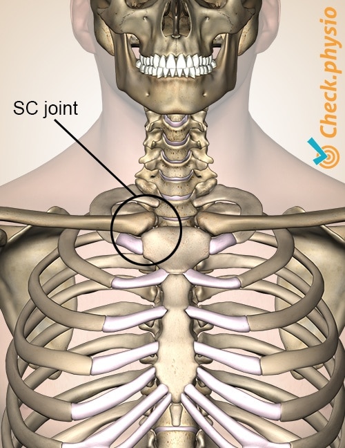
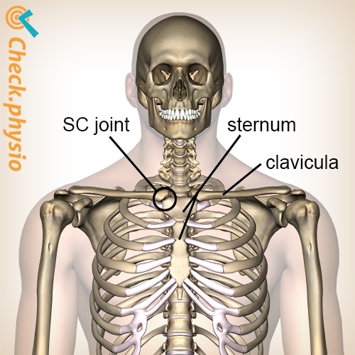
The sternoclavicular joint (SC joint) is located along the front of the chest-neck region. The joint connects the collarbone (clavicle) to the breastbone (sternum). The symptoms usually occur as the result of an accident or fall.

Injury to the sternoclavicular joint is less common than acromioclavicular injury, which is located more to the outside and top of the shoulder.
Description of the condition
In the case of sternoclavicular luxation, there is a dislocation of the collarbone (the clavicle) with respect to the breastbone (the sternum). This causes damage to the structures that keep the joint together. These are primarily capsules and ligaments. Sometimes the disc that is (almost always) located in the joint can also become damaged.
Dislocation does not always have to occur. Several underlying processes can result in pain of the sternoclavicular joint. For example, capsulitis (inflammation of the joint capsule) or arthritis (inflammation of the joint). This is seen primarily in middle-aged women.
Cause and origin
Generally, dislocation will occur as the result of a direct or indirect force acting on the shoulder. For example, an accident or fall. Sometimes the patient cannot remember a specific accident or fall and the symptoms develop gradually.
Signs & symptoms
- Pain along the front of the chest, just below the base of the neck.
- Various fully extended movements can make the symptoms worse.
- Stretching the shoulders or moving the upper arm towards the chest (horizontal adduction) is often painful.
- Pressure on the sternoclavicular joint is painful.
- Localised swelling can occur.
- If dislocation occurs, there can be visible abnormalities in the joint.
- Some patients will complain of shortness of breath, pins and needles, or difficulty swallowing.
Diagnosis
Patients can present themselves to a doctor or physiotherapist at both an acute and chronic stage. In order to find out what the problem is, the patient will be asked what the complaints are and how they arose. The physical examination will reveal any limitations.
An X-ray can show whether there is damage to the joint. However, an X-ray is usually not sufficient in case of a luxation. A CT-scan will give a better indication. An ultrasound could show capsulitis or arthritis.
Treatment
The treatment and the corresponding recovery depend on the nature and severity of the injury. In case of an acute luxation, the collarbone will be put back in place and a Mitella/sling will be worn for a few weeks after that.
If it is not possible to reposition the joint, surgery will be performed. Afterwards, it may be necessary to regain the mobility of the shoulder girdle by means of physiotherapy.
In case of chronic or recurring luxations, it is initially attempted to reduce the symptoms with rest, physiotherapy and, if necessary, painkillers. If the symptoms persist, surgery can also be performed to fix the joint.
In case of a sprain, capsulitis or arthritis of the joint, a period of rest will usually be sufficient.
Exercises
Follow the specially compiled exercise programme with exercises for Sternoclavicular injury here.
You can check your symptoms using the online physiotherapy check or make an appointment with a physiotherapy practice in your area.

References
Brukner, P. & Khan, K. (2010). Clinical sports medicine. McGraw-Hill: Australia. 3e druk.
Hout, J.A.A.M. van den & List, J.J.J. van der (1994). Sternoclaviculaire luxatie. Ned Tijdschr Geneeskd. 3dec;138(49).
Malik, S., Chiampas, G. & Leonard, H. (2010). Emergent evaluation of injuries to the shoulder, clavicle, and humerus. Emerg Med Clin N Am 2010;28:739-763.
Nugteren, K. van & Winkel, D. (2007). Onderzoek en behandeling van de schouder. Houten: Bohn Stafleu van Loghum.
Verhaar, J.A.N. & Linden, A.J. van der (2005). Orthopedie. Houten: Bohn Stafleu van Loghum.
Wolf, A.N. & Mens, J.M.A. (2001). Onderzoek van het bewegingsapparaat. Fysische diagnostiek in de algemene praktijk. 3e, geheel herziende druk. Houten: Bohn Stafleu van Loghum.


